Abdominal Blood Vessels Labeled / Wire Models : The blood vessels are the components of the circulatory system that transport blood throughout the human body.
Abdominal Blood Vessels Labeled / Wire Models : The blood vessels are the components of the circulatory system that transport blood throughout the human body.. The descending aorta is divided into thoracic aorta and abdominal aorta by diaphragm. Human anatomy for muscle, reproductive, and skeleton. Blood, the heart and the vessels that carry blood around the body together make up the cardiovascular system. Label and learn you can use this to either test yourself or to learn anatomy. Incidence of abdominal wall defects is related to surface water atrazine and nitrate levels.
Blood vessels of the upper limb. This activity contains 12 questions. Blood, the heart and the vessels that carry blood around the body together make up the cardiovascular system. Blood vessels labeled simple : This paper presents an automated anatomical labeling method of abdominal arteries.
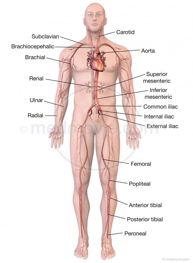
Major Arteries Of The Body Medmovie Com from medmovie.com This paper presents an automated anatomical labeling method of abdominal arteries. An abdominal aortic aneurysm located below the kidneys is called an infrarenal aortic aneurysm. The intestines have very rich blood supply. Incidence of abdominal wall defects is related to surface water atrazine and nitrate levels. Posterior abdominal wall and blood vessels. Role of the use of omental flap in prognosis of cases with induced acute pancreatitis in. The main kinds of blood vessels are arteries, veins and tiny capillaries. Between arteries and veins, there is a network of.
The thoracic aorta supplies blood to viscera of the.
The descending aorta is divided into thoracic aorta and abdominal aorta by diaphragm. Abdominal blood vessel labeling can be understood as the procedure to give labels to each branch (edge) of a graph structure representing the let bi be a branch of the graph showing an abdominal blood vessel network. The blood vessels make up the body's cardiovascular system. The abdominal aorta is the largest blood vessel in the abdomen. This activity contains 12 questions. Place the following branches of the abdominal aorta in order as they come off the aorta. Dimitrios mytilinaios md, phd • last reviewed: Blood vessels are vital for the body and play a key role in diabetes helping to transport glucose and insulin. New blood vessel growth is called angiogenesis. Incidence of abdominal wall defects is related to surface water atrazine and nitrate levels. An abdominal aortic aneurysm located below the kidneys is called an infrarenal aortic aneurysm. Human anatomy for muscle, reproductive, and skeleton. Pictures and 3d models played a great role in helping me learn anatomy.
The best websites voted by users. While most blood vessels are located deep from the surface and. Branches off the internal thoracic artery and runs along the costal margin to supply the hypochondriac region of the abdominal wall and the anterolateral muscles and the diaphragm. The blood vessels make up the body's cardiovascular system. Dimitrios mytilinaios md, phd • last reviewed:
Principal Veins from oer2go.org All cells in the body need oxygen and the vital nutrients found in blood. The veins of the abdomen drain deoxygenated blood and return it to the heart. Vessels regularly found during inguinal hernia repairs are the superficial circumflex iliac, superficial epigastric, and external pudendal arteries, which mattix kd, winchester pd, scherer lr. 1) starts at entry into abdominal cavity through aortic hiatus of diaphragm and ends by bifurcating at level l4 vertebrae into right and left common iliac arteries a) runs down midline of abdominal cavity; Label heart and blood vessels. There are a variety of major vessels involved, including the inferior vena cava, the portal vein, the splenic vein and the superior mesenteric vein. Branching pattern of blood vessels differs among individuals. Blood vessels of the upper limb.
Label the veins of the upper limb.
Blood is oxygenated in capillaries that flow through the alveoli of the lungs. Branching pattern of blood vessels differs among individuals. They also take waste and carbon dioxide away from the tissues. The thoracic aorta supplies blood to viscera of the. The blood vessels make up the body's cardiovascular system. Put simply, they are supplied and drained by the branches of three primary vessels: There are a variety of major vessels involved, including the inferior vena cava, the portal vein, the splenic vein and the superior mesenteric vein. Label the blood vessels and structures using the hints provided. Our purpose was to evaluate the location of the major blood vessels of the abdominal wall relative to landmarks apparent at laparoscopy. The blood circles the body around and around your whole life. While most blood vessels are located deep from the surface and. The abdominal aorta is the largest blood vessel in the abdomen. For example, new capillaries permeate the muscles of a conditioned athlete.
They are vital for carrying nutrients, oxygen and waste around the body. Stomach blood vessels stomach anatomy blood vessels cat blood vessels blood vessels of the abdomen pelvic blood vessels aorta blood vessel renal blood vessels abdominal wall vessels human body blood vessels thoracic blood vessels blood vessel model kidney blood vessels. The blood vessels of the body form a circle that begins and ends at the heart. Blood is oxygenated in capillaries that flow through the alveoli of the lungs. Abdominal blood vessels labelled on gross anatomy specimen.
Blood Vessels Of Abdomen And Pelvis Anatomy Overview Kenhub from thumbor.kenhub.com Not only do blood vessels carry oxygen and nutrients, they also transport carbon dioxide and waste products away from our cells. Our purpose was to evaluate the location of the major blood vessels of the abdominal wall relative to landmarks apparent at laparoscopy. Posterior abdominal wall and blood vessels. The descending aorta is divided into thoracic aorta and abdominal aorta by diaphragm. Label the veins of the upper limb. They are vital for carrying nutrients, oxygen and waste around the body. Blood vessels labeled simple : The best websites voted by users.
Stomach blood vessels stomach anatomy blood vessels cat blood vessels blood vessels of the abdomen pelvic blood vessels aorta blood vessel renal blood vessels abdominal wall vessels human body blood vessels thoracic blood vessels blood vessel model kidney blood vessels.
The blood vessels make up the body's cardiovascular system. The celiac, superior and inferior. Between arteries and veins, there is a network of. As a medical student, i found anatomy pretty challenging. Label the steps in the homeostatic response to high blood pressure. Label heart and blood vessels. They are vital for carrying nutrients, oxygen and waste around the body. Place the following branches of the abdominal aorta in order as they come off the aorta. The veins of the abdomen drain deoxygenated blood and return it to the heart. For example, new capillaries permeate the muscles of a conditioned athlete. Posterior abdominal wall and blood vessels. The best websites voted by users. Small aneurysms may go completely unnoticed.
Posterior abdominal wall and blood vessels blood vessels labeled. All cells in the body need oxygen and the vital nutrients found in blood.


0 Komentar